What just happened? Another technology that's long been the thing of sci-fi has taken its first steps to becoming a reality: 3D bioprinting complex human organs. The concept of being able to 3D-print entire replica kidneys, hearts, etc. for patients is an exciting one, and researchers have now designed a new type of 3D bioprinter that uses sound, light, and bubbles to bring that dream a little closer.
3D bioprinting has been around for a long time now. It's a slow process that involves the devices printing just a few cells at a time, layer by layer. The problem is that the method can damage cells and lacks the precision required to create perfect replicas of human tissue.
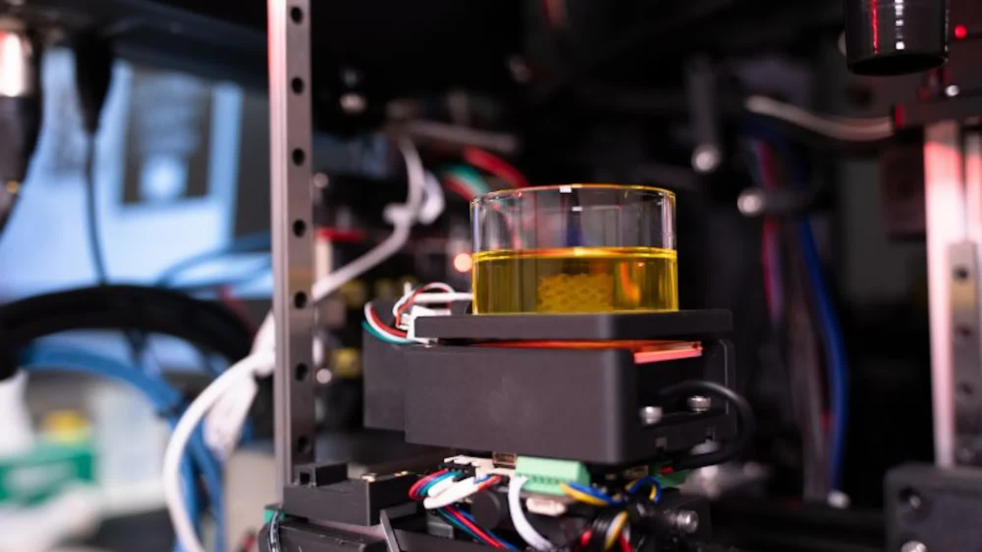
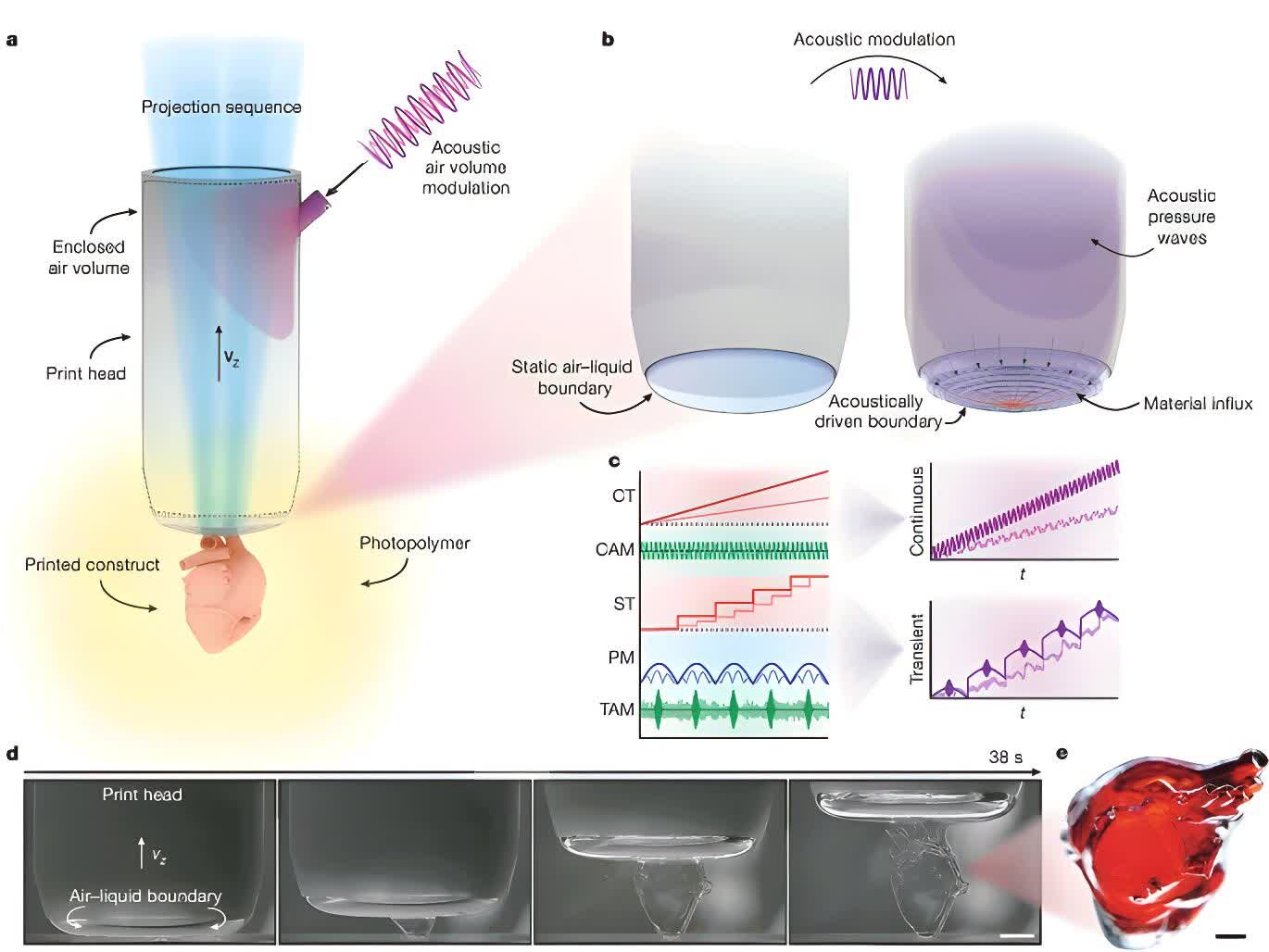
A new process called dynamic interface printing (DIP), created by researchers in Australia, addresses the imprecision problem by printing single cells at a time. It works by filling a tube with liquid polymer then using light to create a bubble and hardening it to match the tissue's shape. Next, a speaker emits sound waves that vibrate the bubble, pushing the 3D-printed cells into place.
Despite the increased precision of the system, it is still more than 350 times faster than traditional 3D bioprinting methods, according to the statement.
"What we're doing is we are shining light in a 2D pattern across – and this is kind of what is the distinguishing feature for this technology – we are printing across a bubble," David Collins, head of the Collins BioMicrosystems Laboratory at the University of Melbourne and a co-author of the study, told ABC Melbourne's Raf Epstein. "We're continuously changing those projections that are curing the individual layers as we go through that. The fundamental principle is that we can shine light onto a material, and we can create a solid."
As the tissue is floating as it is printed, the system can create delicate structures using very soft materials, softer than anything currently being used, according to Collins.
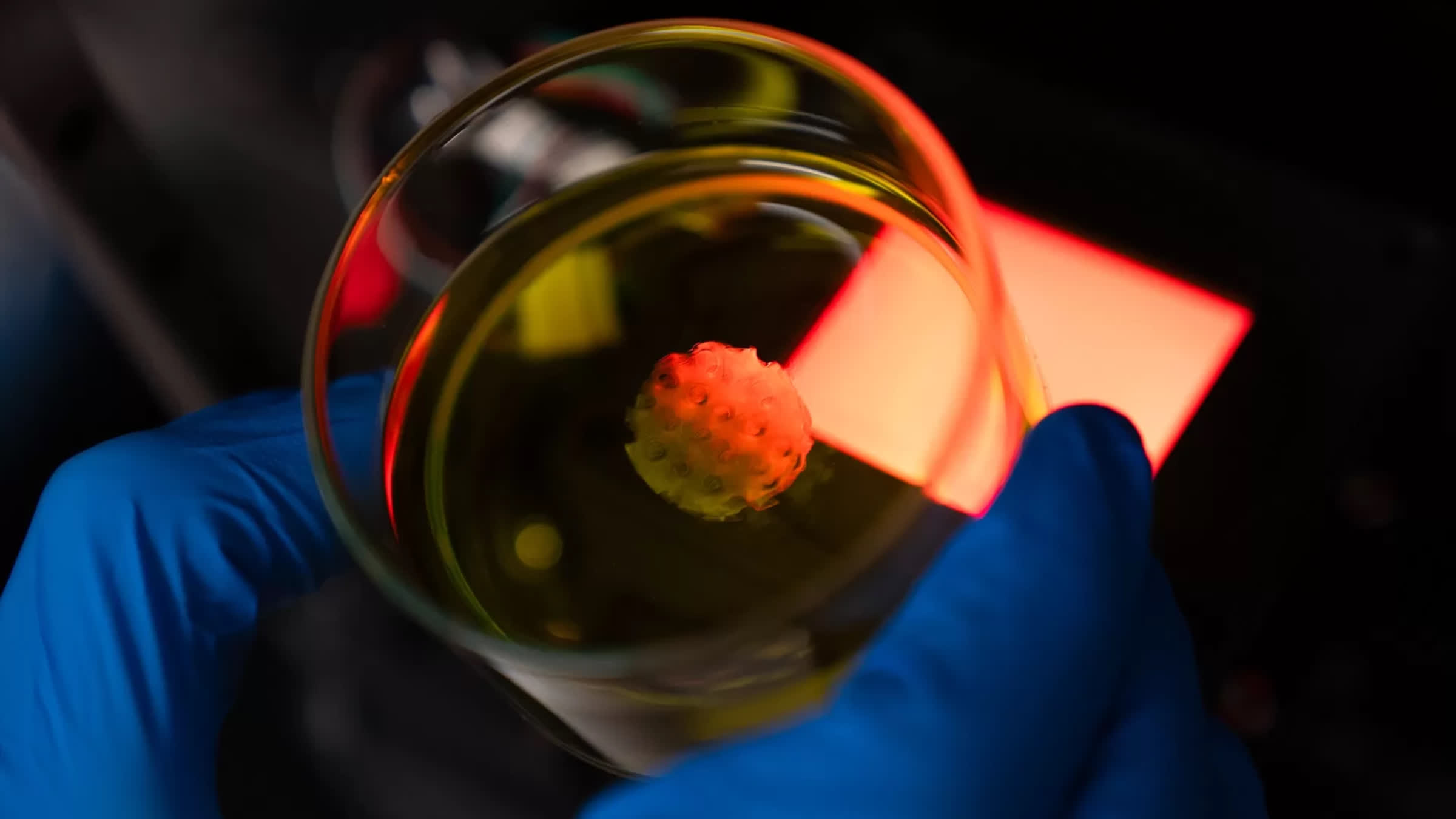
The tissue is also able to be printed directly in a Petri dish rather than the usual hard surface, thereby increasing cell survival rates by eliminating physical handling during tissue transfer. This allows more fragile tissue to be created safely.
The team has so far only printed samples measuring just 3 centimeters in diameter with a length of 7 centimeters and a resolution of 15 micrometers. They envision DIP eventually being used to replicate human organs designed specifically for individual patients or for medical research. It could also replace animal tissue in drug testing trials.
The team said DIP could "help pave the way for more effective, patient-specific therapies in the fight against cancer and other diseases."


